Abstract
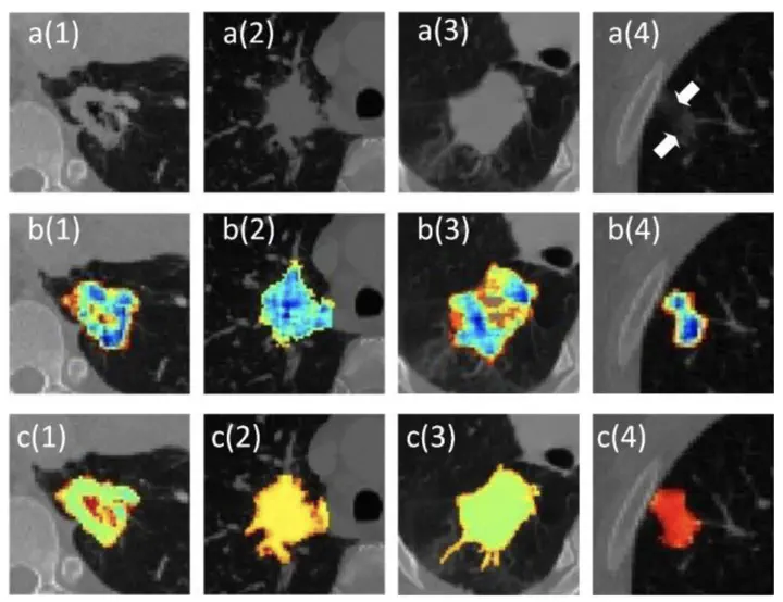
Automatic segmentation of lung nodules on computed tomography (CT) images is challenging owing to the variability of morphology, location, and intensity. In addition, few segmentation methods can capture intra-nodular heterogeneity to assist lung nodule diagnosis. In this study, we propose an end-to-end architecture to perform fully automated segmentation of multiple types of lung nodules and generate intra-nodular heterogeneity images for clinical use. To this end, a hybrid loss is considered by introducing a Faster R-CNN model based on generalized intersection over union loss in generative adversarial network. The Lung Image Database Consortium image collection dataset, comprising 2,635 lung nodules, was combined with 3,200 lung nodules from five hospitals for this study. Compared with manual segmentation by radiologists, the proposed model obtained an average dice coefficient (DC) of 82.05% on the test dataset. Compared with U-net , NoduleNet , nnU-net , and other three models, the proposed method achieved comparable performance on lung nodule segmentation and generated more vivid and valid intra-nodular heterogeneity images, which are beneficial in radiological diagnosis. In an external test of 91 patients from another hospital, the proposed model achieved an average DC of 81.61%. The proposed method effectively addresses the challenges of inevitable human interaction and additional pre-processing procedures in the existing solutions for lung nodule segmentation. In addition, the results show that the intra-nodular heterogeneity images generated by the proposed model are suitable to facilitate lung nodule diagnosis in radiology.